Towards Context-Aware Just-In-Time Information

Micro-Activity recognition of Everyday Objects

  2013


Abstract

Transferring computation tasks from user-worn devices to everyday objects allows the users to focus on their regular non-computing tasks. Identifying micro-activities (short-repetitive-activities that compose the macro-level behaviour) enables the understanding of subtle behavioural changes and providing just-in-time information without explicit user input. In this paper, we propose the concept of micro-activity recognition of augmented-everyday-objects and evaluate the applicability of machine learning algorithms that are previously used for macro-level activity recognition. We outline a few proof-of-concept application scenarios that provides micro-activity-aware just-in-time information.

Introduction

Ubiquitous computing intends to increase the use of computers by making them accessible from anywhere in the physical environment at anytime yet without drawing user’s attention. Recent boom of wearable devices such as smart-watches, smart-glasses attempt to overcome this issue by proposing interactions that require minimal user involvement. However, in instances where people interact with shared physical objects in common spaces, it is more practical to augment the objects rather than humans. For example, in a library, attaching a sensor to a book is more realistic than having each library user wear a special device. The way we interact with everyday-physical-object consists of sequences of short-repetitive-activities which we refer to as micro-activities. A collection of micro-activities make up one macro level activity, which is typically tested in activity recognition research. For instance, cycling can be thought of as a sequence of micro-activities such as accelerating, decelerating, turning, standing still, falling, etc. A rapid intermittent acceleration and deceleration trigger a turbulent ride while constant speed triggers a steady one although both situations fall under the category of cycling at macro level. For understanding such delicate behavioral changes of everyday objects, recognition of underlying micro-activities is of much importance.

Read the full paper

Detection of Microfilariae in Peripheral Blood Smears using Image Analysis

  2013


Abstract

Lymphatic filariasis is a leading cause of permanent disability in many countries. Due to its asymptomatic and epidemiological characteristics, the whole population living in threatened areas need to be screened. The popular diagnostic method involves the manual microscopic observation of nocturnal blood smears in order to detect the presence of microfilariae. Due its strenuous and mundane nature, considerable detection errors are observed. This paper presents an image-based technique for diagnosis of filariasis through the detection of microfilariae present in Giemsa and Hematoxylin and Eosin stained peripheral thick blood smears. The proposed method uses connected component analysis and dynamic thresholding to detect microfilariae in images acquired from the microscope eyepiece. A sensitivity of 91.42% and a specificity of 88.57% are observed on experiments conducted on a database of 70 images

Introduction

Lymphatic Filariasis (LF) is a neglected tropical disease and a leading cause of permanent disability in many countries. By 2013, nearly 1.4 billion people in 73 countries are threatened by LF while about 120 million are currently infected and 40 million are disfigured and incapacitated. About 65% of the infected population resides in the Southeast Asia and another 30% resides in Africa. LF is a parasitic disease caused by the nematodes belonging to the Filarioidea superfamily. Adult worms of three types; Wuchereria bancrofti, Brugia malayi and Brugia timori can cause LF through occupancy in the human lymphatics. Millions of Microfilariae (MF) will be released in to the blood stream of the patient during the 6-8 year lifespan of the adult worm. LF is transmitted through vector borne transmission and the prominent vectors are mosquito species, Culex, Anopheles and Aedes. Most patients will remain asymptomatic carriers, and therefore, mass drug administration is the World Health Organization (WHO) recommendation for threatened areas. Disease status is monitored through nocturnal mass blood testing.

Read the full paper

Automated Whole Slide Imaging

  2013


Abstract

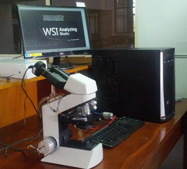
Optical microscopes are used to observe magnified views of specimens that are infinitesimal to be directly observed by the human eye. They are commonly used in histopathology, where urgent reports are often crucial in decision-making. How- ever, dependance on microscope technicians has several identified drawbacks, such as, the field of view being only available to the technician and continuous observation of slides inducing human errors. Furthermore, prolonged use of microscopes can lead to severe eye injuries. To address these issues, Whole Slide Imaging (WSI) microscopes, capable of automatically digitizing slides, are available in the market with a high price tag. However, these sys- tems do not provide the user with the ability to change lenses and it is well known that some users have specific brand preferences in microscope lenses and systems. As a solution, this paper presents a method to enhance the functionality of a conventional optical microscope into a WSI microscope by motorizing the navigator and the fine-tune knob. An eyepiece camera is used to obtain photographs synchronously at predefined positions. The obtained images are then registered and stitched to generate a WSI, directly comparable with the output from the microscope. These images have various applications, such as, teaching, collaborative decision making and computer-aided-diagnostics.

Introduction

Conventional optical microscopes add great value to many applications in the field of medicine. Microscopes are man- ually operated by trained medical laboratory technicians (MLTs). The specimen slide is first placed between the stage clips. Inspection is then carried out through the eyepiece by moving the slide under the objective lens using the navigator. The image is focussed using the coarse and fine tune knobs, located on the body of the microscope. However, such microscope systems have many identified drawbacks. The field of view is limited to the MLT, which means that the accuracy of the analysis is dependent on the alertness, thoroughness and the capability of a single person [1]. Therefore, in order to maintain the consistency, laboratories need to routinely assess the performance of MLTs, which is a difficult task amidst heavy workloads. Even highly skilled MLTs are bound to cause errors when a large number of repeated observations are required [2], [3]. Furthermore, long term microscope usage is an identified ergonomic hazard that can result in traumatic eye injuries and psychological disor- ders. All these complications will lead to further degradations of the output [4]. Read the full paper

Read the full paper